

Where do pigment cells come from?
Neural Crest
Dogs have pigment cells in their skin and hair follicles.
These melanocytes start their journey very early during puppy development.
They originate from a temporary structure in the embryo called the neural crest. This is a population of migratory stem cells in the area that will eventually develop into the brain and spinal cord.
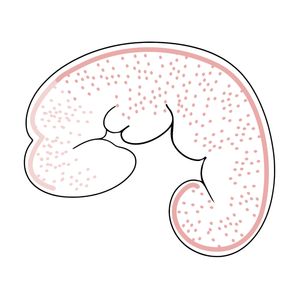
At this early stage, you can vaguely see where the head and the body will be. The neural crest-derived stem cells migrate to different parts of the embryo depending on where they originated.
Each stem cell can differentiate into a wide variety of tissues. Depending on their future job, they migrate outward along defined pathways to many different destinations in the embryo.
Once settled in, they complete the differentiation process. For example, they can become facial bone, cartilage, muscle, adipose tissue, eye tissue, parts of the nervous system or … pigment cells.
Migration
Only the mature, fully functional cells that produce melanin are called melanocytes.
Their immature precursor cells that derive from the neural crest are called melanoblasts.
A melanoblast is committed to later become a pigment cell.
But first, each melanoblast has to travel across the body, find its destination, mature into a pigment cell, proliferate and populate a segment of the body with its progeny.
Many pigment cells travel into the epidermis and become skin pigment cells. Others move into the developing hair follicles and will later provide pigment meant for the growing hair shafts.

And some melanoblasts become dormant melanocyte stem cells, providing a continuous, self-renewing, lifelong supply of new pigment cells for tissue repair and to replenish old pigment cells.
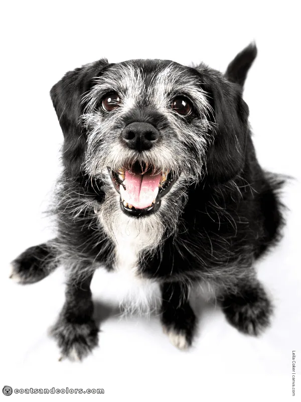
Failure of this self-renewal system is what causes muzzle graying in old dogs.
Pigment cells also colonize other tissues. Dogs obviously need functional pigment cells to produce eye colors. And the inner ear also relies on pigment cells to maintain proper hearing ability.
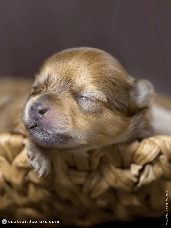
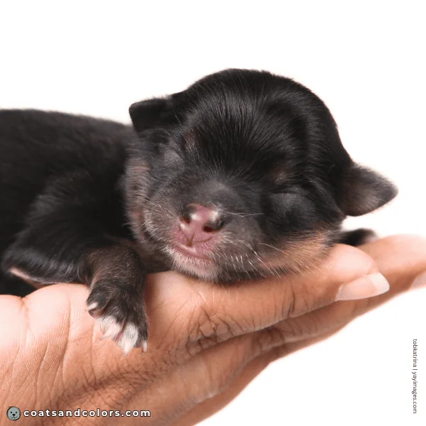
The pigmentation process is still ongoing in newborn puppies. They are born deaf and blind, but their senses all develop rapidly. Many puppies still have weak nose pigment or small white patches of hair that will fill in over time. Even eye colors use to darken in young dogs, which often takes months.
Migration Pathways
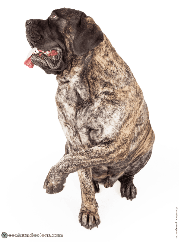
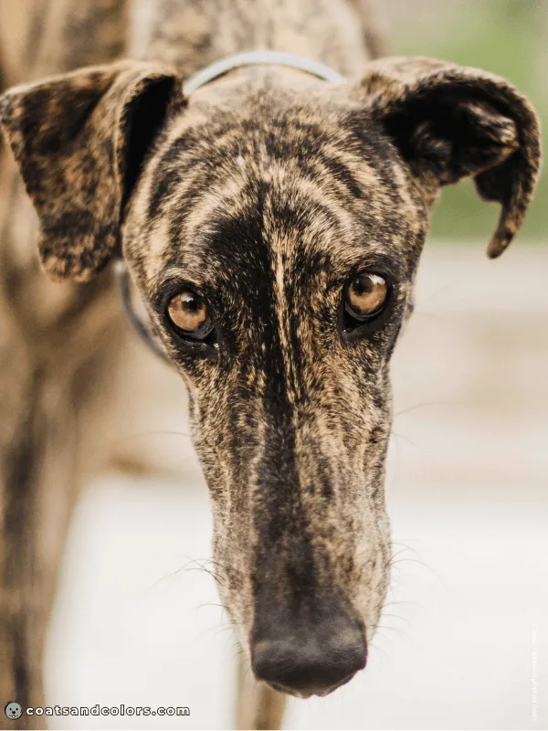
To roughly follow the paths the melanoblasts take, you can trace the stripes on a brindle dog.
The brindle trait causes alternating stripes of dark and light hair.
The brindle allele represents an unstable allele that has the genetics for both solid dark (KBlack) and patterned (kyellow) coat. The kbr allele actually means [KB + ky] ended up on the same chromosome.
Seemingly, pigment cells acquire one of the two alternative “settings” (KB or ky) by chance.
1) Some cells express as ky and show the A locus pattern (e.g. sable).
2) Some cells express as KB and replace the pattern with solid dark hairs.

Each cell maintains its behavior via some epigenetic mechanism and passes it down to its clones, so each black stripe on a brindle dog maps out the migration pathway of this family of cells.
This difference in gene expression is a type of functional mosaicism.
Brindle dogs still often have broken stripes. Or so many stripes that you can’t make out where they begin or end. But for most dogs, it works well enough to roughly follow the path their cells took.

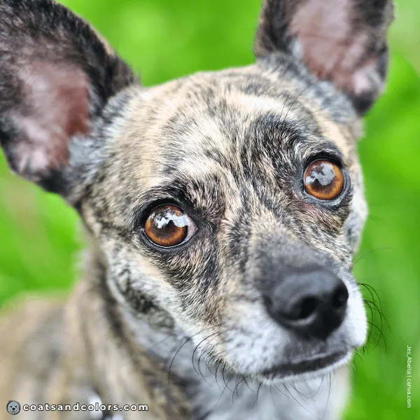
If you follow the black stripes, you will find slightly curved or vertical stripes down the chest, trunk, and legs. And a V-shape down the midline seam, where cells migrated either to the right or to the left.
Stripes reflect the directional outgrowth and clonal expansion of young pigment cells.
Midline Splits
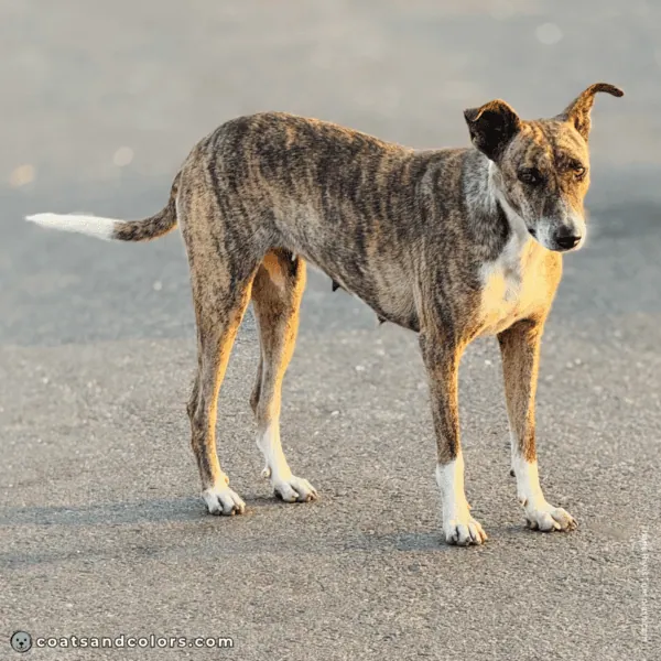
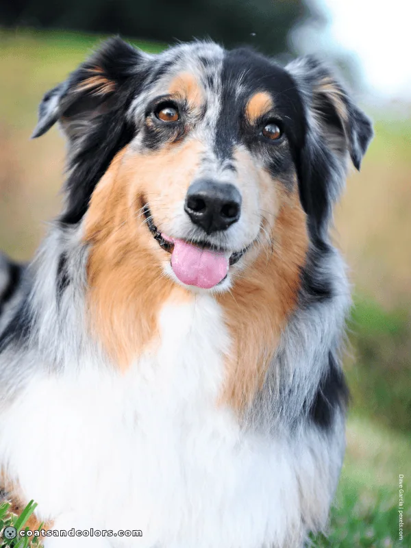
Patterns like brindle are midline-sensitive and can cause a midline split in the visible pattern. This can happen whenever a trait allows for alternative outcomes (e.g. brindle, white, merle).
This boils down to “stochastic events,” meaning it is ruled by chance.
With brindle, cells on one half of the body might behave differently than those on the other half. And white or merle might only disturb pigment cells on one side of the midline, often causing a split face.
White Markings
There is a complex genetic network that regulates early melanoblast formation from the neural crest, cell migration, survival, differentiation into a mature pigment cell, and cell multiplication.
Defects in any one or a combination of the stages cause a lack of living pigment cells.
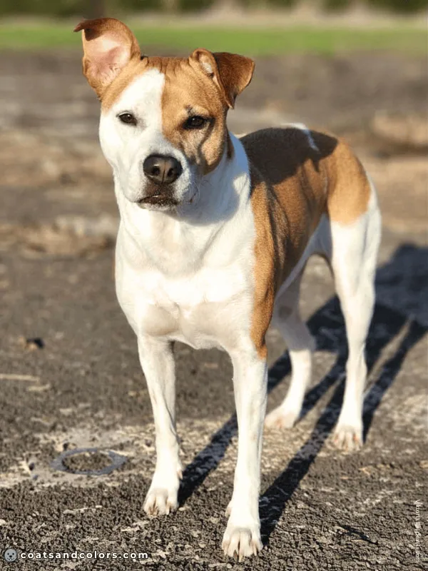
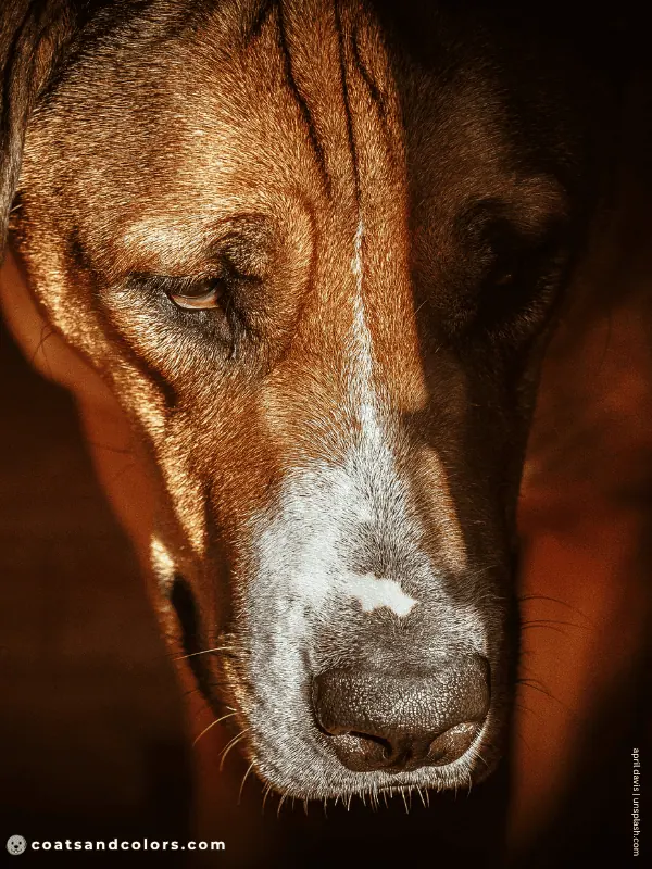
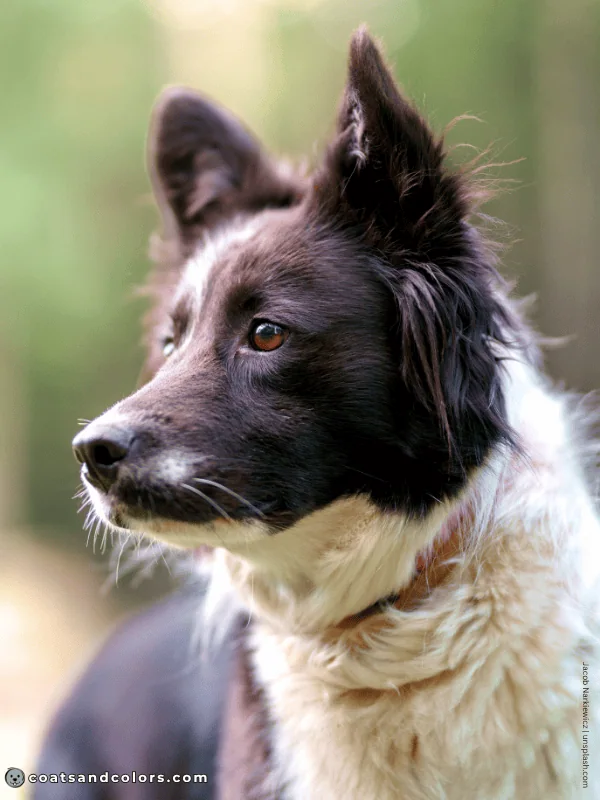
The S locus and other white marking genes do exactly that: They disrupt the pigment cell journey at some point. And if pigment cells fail to colonize an area, this area will have no pigment.
This is how pink skin, blue eyes, or white markings exist in the first place.
Important: A lack of melanocytes in the inner ear can lead to hearing impairment.
Having pigment over the ears can not guarantee the dog has full hearing!
Just as pigment over the eyes does not mean the dog will have brown eyes.
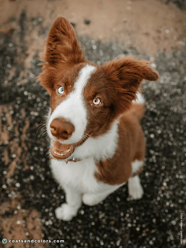
Extended white markings on the face just indicate that pigment cell survival was affected.
It’s just that whatever disturbs skin pigmentation can also impact pigment in the inner ear or delete eye color to blue. The more white on the head, the more likely the dog is also deaf or has a blue eye.
There are still plenty of extreme white dogs with brown eyes and full hearing.
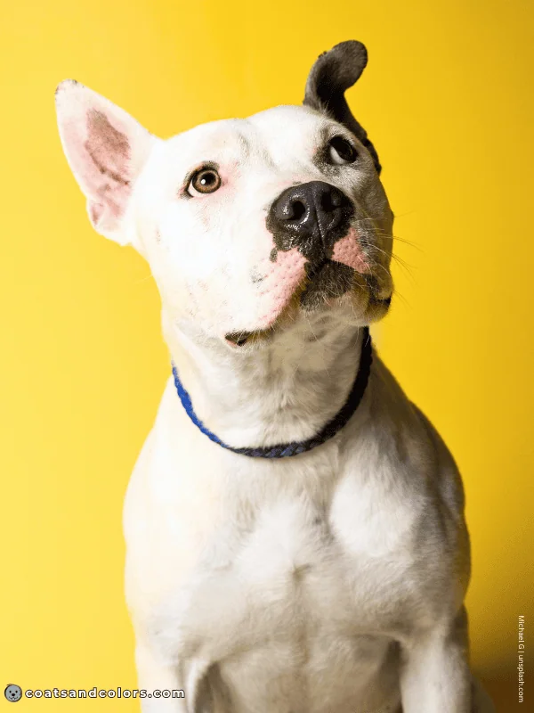
Different white marking traits affect different body parts or maybe different stages of the melanocyte journey. And the amount of white is modified by unknown genetic factors.
This is why each trait can present with a minimal, moderate, or extreme version. And it’s also why the progression of white across the body differs between separate traits.
For example, piebald affects the pigment on the body before it affects the pigment on the head. And whitehead will delete more pigment from the front of the dog before it affects the trunk.
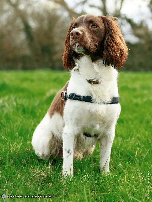
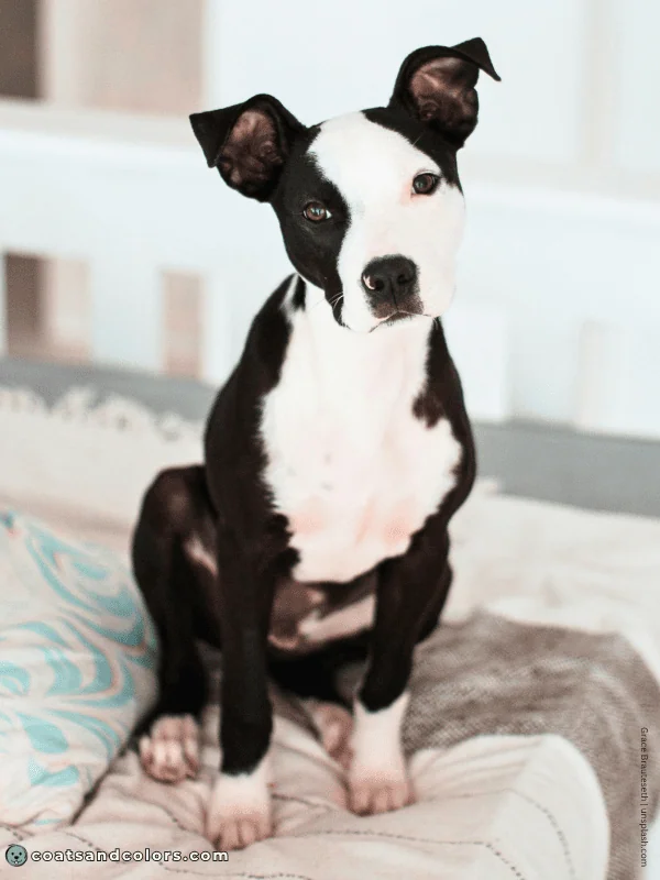
Puppies still build up pigment over time. Some smaller white markings that were visible at birth can fully disappear in the adult coat. And larger white markings can shrink a little around the edges.
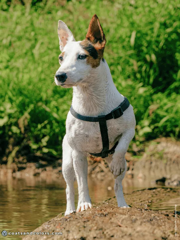
We can also see that some areas are better than others to keep pigmentation. For example, the last areas to lose pigment from piebald are over the eyes/ears and just above the tail. These are probably the areas that get the largest numbers of precursor cells, making it more likely that some of them survive.
The end.
I hope it makes sense now how pigment cell migration can affect certain coat colors in dogs.
Learn More




Links
Kerns et al (2007): Linkage and segregation analysis of black and brindle coat color in domestic dogs. Genetics. doi.org/10.1534/genetics.107.074237
Image Credits
Ákos Nemes/unsplash.com
april davis/unsplash.com
Cavan/depositphotos.com
Celyn Bowen/nsplash.com
Dave Garcia/pexels.com
dpcrestock/yaimages.com
Elina Volkova/pexels.com
Grace Brauteseth/unsplash.com
Jacob Narkiewicz/unsplash.com
Jarrycz/canva.com
Jes_Abeita /canva.com
Korneeva_Kristina/canva.com
Leila Coker/canva.com
Michael G/unsplash.com
Nora Mazzini/canva.com
Thaha Riyas/unsplash.com
tobkatrina/yayimages.com
Viktoria B./pexels.com
Hi! I’m Steffi. I am a biologist and a big time dog nerd. You are curious about coat color genetics? You’ve come to the right place! Read more.